BLOC at 2022 BIST Conference on Precision Medicine

From 8th to 11st of November, BLOC project was present at the 2022 Barcelona Institute of Science and Technology (BIST) Conference on Precision Medicine. Researchers from the “Molecular Imaging for Precision Medicine” laboratory at IBEC presented scientific advances and participated in an outreach event with high school students.

The Barcelona Institute of Science and Technology (BIST) is a leading institution of multidisciplinary research encompassing seven Catalan research centres of excellence, including IBEC, coordinator of BLOC project. In the frame of the 6th edition of the BIST Conference, during 4 days more than 250 people from the BIST Community and its stakeholders shared their research advancements in precision medicine from multiple perspectives and approaches. Precision medicine involves patient-centred and comprehensive approaches to face healthcare, incorporating individual’s genetics, environment, and lifestyle viewpoints into targeted disease prevention and treatment plans.

In this scenario, Irene Marco-Rius presented the work being done in her laboratory at IBEC, that aim to develop molecular imaging tools to identify abnormalities of cell metabolism in human disease and predict treatment efficacy. She centred her talk on the subject of BLOC project and on use of NMR methods to identify biomarkers to diagnose, stage and evaluate treatment response in several diseases, in real time using so-called Hyperpolarization MR techniques.

On his side, Marc Azagra presented a flash talk entitled “High-throughput analysis in a microfluidic multiwell plate by non-invasive dynamic nuclear polarization-magnetic resonance”. He focused on the use of this high sensitivity technique, that is able to enhance the intrinsically low sensitivity of NMR up to 10,000 times allowing its extrapolation for real-time in-situ metabolomic analysis and its potential use as a preclinical tool.

Finally, Alba Herrero participated in an outreach activity in the framework of the 2022 “Setmana de la Ciència”, tailored to high school students. In total, about 2000 students could get in contact with precision medicine research projects explained in an easy way by PhD students. Alba gave an online talk and explained what “molecular imaging” is and how it can be applied to the field of precision medicine. 

BLOC researchers at IDIBAPS receives the Diabetes Association of Catalonia

Anna Novials and Vincent Ribes presented their research to members of the Diabetes Association of Catalonia. The visitors had the opportunity to learn a bit more about the disease and to know the cutting-edge research that is being carried out at IDIBAPS.

In the frame of a new communication project from IDIBAPS, researchers from BLOC Project at the Diabetes Pathogenesis and Prevention group received patients and their families from the Diabetes Association of Catalonia (ADC). ADC is a non-profit organization that supports research projects to develop new treatments, and why not to find a cure for diabetes. They give assistance to more than 600,000 people with the disease in Catalonia.

Anna Novials and Vincent Ribes showed IDIBAPS’s last advances in the field of diabetes research. Among the topics, they explained the BLOC Project and its objectives, aiming to develop a benchtop device based on NMR to detect and monitor metabolic diseases as diabetes to advance towards finding solutions and treatments to improve the life quality of patients. 

Visitors also had the opportunity to find out the cellular models with which the scientists work and to see the equipment that is used to analyse how exercise induces metabolic and molecular changes in patients with type 1 and 2 diabetes.

Sources:

https://www.clinicbarcelona.org/en/news/idibaps-receives-representatives-of-the-diabetes-association-of-catalonia

https://www.clinicbarcelona.org/en/news/representatives-of-the-diabetes-association-of-catalonia-visit-idibaps

One more year BLOC is present at the IBEC Annual Symposium

Last 19th of October it took place the 15th edition of the IBEC Symposium, an annual meeting where bioengineering is in the centre of the scene.  Among several renowned national and international invited researchers, BLOC was present with a flash talk and a poster showing part of the work done at the “Molecular Imaging for Precision Medicine” laboratory at IBEC.

The 15th IBEC Symposium was, in the words of IBEC’s director Josep Samitier, “a great opportunity to share our science, make new connections and also reconnect”. About 250 assistants joined together to present their research and discuss about how bioengineering can contribute to improve the life quality of society.  

In this scenario Marc Azagra presented a flash talk about the use of hight-throughput dynamic nuclear polarization (DNP)-nuclear magnetic resonance (NMR) imaging for real-time and non-invasive metabolic studies using microfluidic multiwell devices. The proposed method represents a new approach for hyperpolarization-enhance NMR experimentation that can be applied in the future as a preclinical tool to study diseases that affect cellular metabolism.

Additionally, Lluis Mangas presented a poster on the use of hyperpolarized nuclear magnetic resonance to study hepatocellular metabolism as part of his work in BLOC Project. Concretely, he focused on the development of a MR-compatible bioreactor platform for hepatocellular carcinoma metabolic analysis in real-time using a 60 MHz benchtop NMR spectrometer.

“Gym on a chip”: a new tool to help fighting against diabetes

Researchers led by Javier Ramón-Azcón from the Institute for Bioengineering of Catalonia develop a “gym on a chip” that will help to study diabetes and find new drugs to treat the disease. The dispositive allow to study, in vivo, the crosstalk among different organs. In this paper, researchers combine muscle and pancreatic cells on a single chip and demonstrates that insulin production by the pancreas during exercise is induced by the contraction of muscle cells.

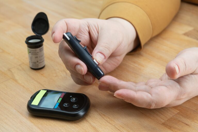
According to the World Health Organisation, 422 million people worldwide suffer from diabetes mellitus, and 1.5 million deaths each year are directly associated with the disease. Diabetes is a complex metabolic disease characterised by a high accumulation of glucose in the blood resulting from a failure in the production or activity of insulin. Insulin, in turn, is a hormone produced, stored and released by the pancreas, which enables glucose to enter the different cells of the body to provide them with the energy they need to function properly. Muscle is one of the main targets of insulin and is crucial in the overall maintenance of glucose levels throughout the body.

The development of new drugs to prevent and treat diabetes must be based on tools that take into account the communication between the pancreas and other organs in order to recreate the details of the disease. In a recent study published in the Advanced Materials Technology journal, IBEC researchers led by Juan M. Fernández-Costa and Javier Ramon-Azcon from the “Biosensors for Bioengineering” group, have developed a “multi-organ-on-chip” that allows, in a single device, to study the communication between the insulin-producing cells of the pancreas and the muscle cells. By using this innovative “gym on a chip”, researchers found that muscle cells contraction caused by electric stimulation directly induces the production of insulin by pancreatic cells.

The “gym on a chip”

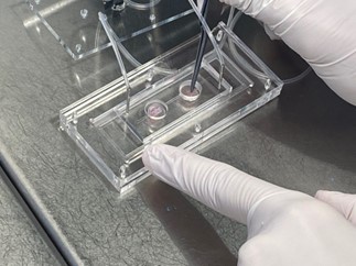
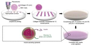
The “multi-organ-on-chip” device engineered by the researchers have one compartment with skeletal muscle cells and another one with pancreatic islets composed of beta cells, which produce insulin. As these cells grow and develop, they form 3D structures that simulate a “mini-organ” inside the chip.

The muscle cells of the “multi-organ-on-chip” are connected to an electrical stimulator that simulates physical exercise and induces their contraction. In addition, the device is also integrated with a complex biosensor platform capable of monitoring insulin levels produced by pancreatic cells, and interleukin-6 (IL-6) levels without the need of external labelling. IL-6 is a regulatory protein that is produced by the muscle and signals the pancreas to increase insulin production. In other words, during exercise, IL-6 moves from the muscle to the pancreas to stimulate insulin production.

“With our “gym on a chip” we demonstrate that insulin secretion by pancreatic islets during exercise is dynamically mediated by the contractile activity of muscle cells and not by other intermediates”. Juan M. Fernández-Costa, first author of the study.

The new chip is a powerful tool for diabetes drug development and testing, and it allows personalised modelling of the disease by using patient’s own cells. With this model, researchers and clinicians will have the possibility to study diabetes following a new approach, by combining different cell types, and study in detail why insulin production by the pancreas sometimes fails.

In the future, the “muti-organ-on-chip” could also be used to study communication between other organs in the context of various metabolic diseases, providing a promising platform for new drug screening and personalised medicine.

Reference article: Training-on-a-Chip: A Multi-Organ Device to Study the Effect of Muscle Exercise on Insulin Secretion in Vitro. Juan M. Fernández-Costa, María A. Ortega, Júlia Rodríguez-Comas, Gerardo Lopez-Muñoz, Jose Yeste, Lluís Mangas-Florencio, Miriam Fernández-González, Eduard Martin-Lasierra, Ainoa Tejedera-Villafranca, and Javier Ramon-Azcon. Adv. Mater. Technol. 2022, 2200873.

A cryopreservation method for bioengineered 3D cell culture models

Alba Herrero, Marc Azagra and Irene Marco-Rius from the Molecular Imaging for Precision Medicine Laboratory at IBEC, have developed a viable cryopreservation method to preserve bioengineered, three-dimensional (3D) cell models, including patients’ samples. The results of their work have been recently published on the Biomedical Materials Journal.

Cryopreservation of biological material as living tissue, cell lines and primary cells is a technique widely used in most molecular biology laboratories around the world. However, this technology was not adapted for its use on three-dimensional (3D) cell models. What Bloc researchers, led by Irene Marco-Rius, coordinator of BLOC project, have done now is to go a step further in this field and develop a viable protocol for spheroid cryopreservation and survival based on a 3D carboxymethyl cellulose scaffold and precise conditions for freezing and thawing.

By applying the new cryopreservation protocol, researchers were able to efficiently cryopreserve bioengineered hepatocytes, for which the scaffold provides both the 3D structure for cells to self-arrange into spheroids and to support cells during freezing for optimal post-thaw viability.

“The use of the 1% Carboxymethyl cellulose cryogel scaffold is crucial to procure support for the cells through the process as well as to aid in the removal of the cryoprotective agent after thawing for improved cell viability”.

This new protocol could be adapted for other bioengineered tissues and may be used to applications that demand high-integrity preservation and transport of pre-assembled 3D models, as cell lines and samples from patients. Moreover, the methodology described in this work pave the way for the development of protocols that simplify and reduce the cost and infrastructures required for the research with 3D cell cultures.

Reference article: Alba Herrero-Gómez, Marc Azagra, Irene Marco-Rius. A cryopreservation method for bioengineered 3D cell culture models. 2022 Biomed. Mater. 17 045023. DOI 10.1088/1748-605X/ac76fb

BLOC experts: interview with Matthew Fallon

BLoC is a European Project that brings together different academic and industrial partners to improve a new technology that integrates tissue engineering and magnetic resonance spectroscopy using dynamic nuclear polarisation (DNP-MR), to monitor diabetes and liver diseases.

Matthew Fallon is a mechanical engineer with experience in several engineering fields as draughtsperson, CAD engineer and project engineer. Few months ago, he joined BLOC Project, where he brings all his knowhow working as a junior designer at Oxford Instruments. For sure an important piece for the project!

Can you describe yourself in a couple of lines?

I am originally from a small town in county of Herefordshire. I lived there until I was 18 years old to which I moved to Plymouth to study a bachelor’s in mechanical engineering. After I graduated with honours I got a job as a draughtsperson in a steam engineering company in Cheltenham for a year and a half. I moved over to Surrey to work for a generator installation company as a CAD engineer and then as a project engineer. Finally, I have now moved over to Oxford to work for Oxford Instruments as a Junior Designer.

What is your role/position within BLOC?

I am currently designing and testing the carrier for the chip, which shall be placed into our X-Pulse system.

Could you tell us a little bit about the concrete work you are involved in inside BLOC project? 

I have been working on prototype versions of the carrier for the chip. These are mainly for fit and function testing. This gives us a good idea of what the carrier will look like, how it will fit into the X-Pulse, and how well the chip will be held. I am currently researching the best and most cost-effective techniques that can be used to manufacture the carrier.

What are the expected results?

To have a cost effective, strong, reliable method for holding the chip inside our X-Pulse. The carrier will have to be highly accurate and position the chip in the same place every time.

What is the expected impact of the work you’re doing?

To allow researchers to have an easy to use and reliable method of placing the chip inside the X-Pulse. This will allow for accurate measurements and the best possible chance of a successful experiment. The design shall be robust and work in such a way that it cannot be done wrong.

How do you feel about being a part of this European Project?

This is a great opportunity to learn and develop my skills as an engineer. It is very exciting to see how other countries and professionals work together and develop a future product.


The other members of the BLOC Consortium are:  IBECMultiwave Technologies and IDIBAPS.
If you want to know more about the project check the BLOC webpage.


Innovative bioengineered spheres might help treating diabetes

Researchers led by Javier Ramón from BLOC project, in collaboration with IDIBAPS research center in Barcelona, have developed nontoxic small spheres able to respond to variations in glucose levels, and producing insulin in vitro. These biomimetic spheroids containing pancreatic β-cells were prepared based on 3D bioprinting. This approach might help in the future improving clinical outcomes of β-cell transplantation strategies for diabetes treatment, as well as for in vitro drug screening platforms.

Diabetes Mellitus is a metabolic disease characterized by a failure in the production of insulin by the β-cells in the pancreas, the hormone responsible to regulate glucose levels in the blood. In 2019, according to the World Health Organization, diabetes caused the death of 1.5 million people worldwide, and nowadays, there are more than 420 million people affected by this disease.

In type 1 diabetes (T1DM), an autoimmune disease, a type of cells called β-cells and found in the pancreatic islets, are destroyed by the immune system, avoiding the production of insulin. The most extended treatment in this case is the regular injection of insulin by the patients, throughout all their lives, to control the persistent high glucose levels in the blood. However, as this therapy does not simulate the real time secretion of insulin by the β-cells, it ends by causing severe chronic complications, as cardiovascular diseases and nephropathy.

A promising alternative therapy against T1DM is the transplantation of pancreatic islets containing β-cells to the patients. Unfortunately, it cannot be currently applied in the clinic due to complications as the poor oxygenation and vascularization of the cells, that highly limit their retention following implantation.

To overcome these obstacles, Javier Ramón, BLOC researcher at Institute for Bioengineering of Catalonia (IBEC), led a work in collaboration with researchers from IDIBAPS, to develop a high-throughput methodology applying 3D bioprinting to encapsulate β-cells inside collagen-tannic acid spheroids. This innovative strategy can increase the success of pancreatic islet transplantation and has been recently published in the Advanced Materials Technologies Journal.

3D bioprinted spheroids containing β-cells produce insulin in vitro 

One strategy to increase β-cells retention after transplantation is to use protective semipermeable biomaterials to encapsulate the cells. These engineered tissues, known as spheroids or encapsulated-based microspheres, provide biocompatible properties that confer a more effective cell attachment, thus reducing cell loss, and acts as a physical protective barrier against the patient’s immune system.

The new approach proposed by IBEC researchers employs 3D bioprinting technology to develop collagen spheroids crosslinked with tannic acid, in an attempt to mimic the extracellular matrix microenvironment of in vivo β-cells. Crosslinking with tannic acid prevents collagenase degradation, enhances spherical structural consistency, and allows customization of microsphere diameter with extremely low variability

To validate the new strategy, researchers prepared 3D collagen-based microcapsules crosslinked with tannic acid containing rat insulinoma cells. They observed that the spheroids maintained cell viability and metabolic activity up to 30 days, reflecting a correct diffusion of oxygen and nutrients. Also, the tannic acid crosslinking strategy improved the retention of β-cells inside the spheroids.

“Our high-throughput approach facilitates the fabrication of a cell encapsulation system that ensures good survival and functionality of insulin-producing β-cells in a glucose-based manner”

This novel 3D bioprinting procedure allows the fabrication of a large number of microspheres capable to respond to glucose by secreting insulin and can be helpful for advanced functional studies aiming β-cell transplantation for diabetes treatment. Additionally, this technology can open new avenues and be applied to encapsulate a wide range of transplantable cell types.


Reference article: Laura Clua-Ferré, Francesco de Chiara, Júlia Rodríguez-Comas, Jordi Comelles, Elena Martinez, Amelie Luise Godeau, Ainhoa García-Alamán, Rosa Gasa, and Javier Ramón-Azcón. Collagen-Tannic Acid Spheroids for β-Cell Encapsulation Fabricated Using a 3D Bioprinter. Adv. Mater. Technol. 2022, 2101696.

Unique resonance technology within reach of BLOC researchers

A Dynamic Nuclear Polarisation (DNP) Polariser for Nuclear Magnetic Resonance (NMR) has been recently installed at IBEC, coordinator of BLOC project, being the only one in operation in Spain, and among the few ones existing in Europe. This equipment can improve up to 10000 folds the signal compared to a conventional NMR procedure and allows for the study of cellular metabolism in real time.

The HyperSense™ equipment, from Oxford Instruments Molecular Biotools, has been recently installed in the Institute for Bioengineering of Catalonia (IBEC), at the Molecular Imaging for Precision Medicine laboratory (MIPMED) led by the coordinator of BLOC project, Irene Marco, following a cession agreement made with the University of Barcelona (UB).  This in vitro polarizer can provide a signal-to-noise ratio not reachable by conventional NMR equipment, increasing sensitivity by more than 10,000 fold. As a result, this technique can be used to study rapid metabolic processes either in vivo or in vitro, in a non-invasive way and in real time, providing information on the mechanistic and biochemical changes that occur in a diseased organ, for example. This powerful technique can open new areas of research and will probably change the field of NMR.

The European Project BLOC will strongly benefit from this recently installed equipment, as this forefront technology is crucial to fulfill its main objective: to develop a benchtop spectrometer based on magnetic resonance spectroscopy and imaging using dynamic nuclear polarisation (DNP-MR) to monitor metabolic diseases such as diabetes and non-alcoholic fatty liver disease (NAFLD) and evaluate responses to different stimulus.

The equipment installed at IBEC performs dynamic nuclear polarisation of samples in the fluid-form, for example sugars, that are afterwards used to monitor cellular metabolism on a conventional NMR equipment. The sample is transferred to the center of the HyperSense™, where it is submerged in liquid helium at the core of the magnetic field. Everything is done under an extremely low temperature, -271,75 ºC, which allows the sample carbon atoms to be nearly fully polarised.

BLOC researchers use hyperpolarised pyruvic acid, a sugar that is then used to study the metabolism of different cellular types. Inside the NMR, the cells are supplied with this sample and, for about 75 seconds, researchers can follow up its absorption by the cells and all the metabolic steps that use this sugar as energy source. This short time is sufficient to see several enzymatic reactions and transient metabolic reaction intermediates. In other words, this remarkable boost allows researchers to see molecular processes of different biological systems in real time, in situ, and in a non-invasive way, in just few minutes. Moreover, as it is a very fast procedure, problems such as temperature and oxygen changes, cell death and cellular alterations during the experiment are avoided, allowing researchers to reliably compare results among different experiments.

The hyperpolarised magnetic resonance technique allows us to observe in a couple of minutes what we could only see in hours with a conventional NMR, and moreover with a sensitivity that can be up to 10000 times higher.

Irene Marco, coordinator of BLOC project

The time-lapse video below shows the entire installation process of the HyperSense™ equipment, which took three days and counted with the participation of various skilled technicians.


BLOC reinforces its commitment to the International Day of Women and Girls in Science

Each 11 February we celebrate worldwide the International Day of Women and Girls in Science with the aim to eliminate gender stereotypes and long-standing biases that keep girls and women away from science. This year, BLOC participated with two talks to 11-12 years-old students to help normalizing the presence of women in scientific fields.

Since 2015, when the United Nations General Assembly declared 11 February as the International Day of Women and Girls in Science, this day is devoted worldwide to achieve full and equal access to and participation in science for women and girls. Through different actions and events, the idea is to further achieve gender equality promoting the empowerment of women and girls and promoting their participation in science.

In this scenario, BLOC contributed to this cause in the frame of the event “100tifiques”, organized by the Catalan Foundation for Research and Innovation (FCRI) and the Barcelona Institute of Science and Technology (BIST), an initiative that aims to bring science closer to schools and claim the female scientific talent around Catalonia.

Alba Herrero, researcher at the “Molecular Imaging for Precision Medicine” laboratory, and Bia Moreno, Communication Officer of the BLOC project, both from IBEC, gave talks to 11-12 years old students from two different schools. They explained her scientific trajectory and the ways that brought them to scientific fields: Alba is graduated in Biochemistry and is now doing her PhD, and Bia has a PhD in Molecular Biology.  Both focused her talks not only on scientific aspects, but also on personal and general aspects of their lives, with the objective to show to girls and boys that, despite being scientists, they also have a “normal” live!   

BLOC experts: interview with Samantha Morón

BLoC is a European Project that brings together different academic and industrial partners to improve a new technology that integrates tissue engineering and magnetic resonance spectroscopy using dynamic nuclear polarisation (DNP-MR), to monitor diabetes and liver diseases.

Dr. Samantha Morón is a biochemist from Barcelona with a solid experience in immunology and metabolic diseases. After finishing her PhD, where she studied organ’s reaction to thermogenic response and obesity, she joined the group of Dr. Anna Novials from IDIBAPS to perform a postdoctoral research in the frame of BLOC. Her knowledge and skills are key to the success of the project!

Studying beta cell morphology by confocal microscopy to assess their functionality.

Can you describe yourself in a couple of lines?

My name is Samantha Morón Ros and I am from Barcelona. I have studied a BSc in Biochemistry and an official master’s degree in biomedicine, both in the University of Barcelona. I participated during these years with a research group focused in immunology and the role of macrophages in the immune response. After that I started my PhD in the Molecular Metabolism and Disease’s group led by Francesc Villarroya where I studied the response of different organs to thermogenic response and obesity and the effect of some fibroblast growth factors, such as FGF15/19. After I finished my PhD in 2020, I joined the research group “Pathogenesis and Prevention of Diabetes” led by Anna Novials as a postdoctoral researcher.

What is your role/position within BLOC?

Due to my experience in animal models and my background as a researcher in metabolic diseases, my role as a postdoctoral scientist is to evaluate the beta cell function in in vivo mice models and to support the validation of DNP-NMR measurements.

Could you tell us a little bit about the concrete work you are involved in inside BLOC project? 

As my other colleagues have answered before, one of the main challenges of this project is to develop bioengineered organs compatible with the DNP-NMR technique.  Moreover, the study of the crosstalk between different bioengineered organs is a goal of the BLOC project as well. Right now, I’m working on in vivo experiments evaluating diabetes, obesity and fatty liver mouse models with the objective to evaluate DNP-NMR measurements. On the other hand, I’m working on in vitro engineered models of pancreatic islets to use them with engineered liver for NMR studies.

What are the expected results?

To validate the results, we obtain with DNP-NMR technique, we need a reliable model of the organs we want to evaluate. We expect to have a physiologically accurate pancreatic and liver model compatible with DNP-NMR technique.  When we have these models, we will have reliable models for studying human metabolic diseases with this promising technology.

How do you feel about being a part of this European Project?

As a biomedical researcher, being part of such a multidisciplinary project with professionals from different parts of Europe is very exciting. All the researchers involved in this BLOC project are helping each other. Also, I was always working in projects focused on biochemistry or biomedicine. Participating in the BLOC, which combines the biomedicine but also the technology (with the NMR) is very interesting and I am learning a lot from my colleagues.


The other members of the BLOC Consortium are: IBECOxford Instruments and Multiwave Technologies.
If you want to know more about the project and the other partners, check the BLOC project webpage.