The coordinator of BLoC project Irene Marco-Rius and the PhD student Marc Azagra recently published an article in ”Investigación y Ciencia” (the Spanish Edition of Scientific American), an online specialized media dedicated to the dissemination of scientific culture. They explain in a very elegant and simple way the different techniques currently used to perform molecular imaging and diagnostic (article in Spanish).
In this article, oriented to a general public, Irene and Marc explain the details of several imaging techniques used in medicine to diagnose, evaluate and treat diseases. They also highlight their importance to analyse the response of patients to certain treatments and how they help clinicians to choose the best medical approach.
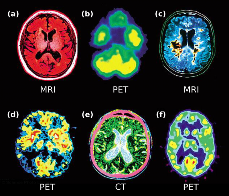
Some techniques as X-rays and derivatives, computed tomography, densitometry and mammography, ultrasound examinations, or optical images are used to give anatomical information about the patient (bones, tumours and ulcers). On the other hand, another group of more recent imaging techniques allow to obtain cellular and molecular information about processes occurring inside our body. Those represent a great advance as a disease can be detected long time before the appearance of anatomical changes. Examples of these techniques are PET (Positron emission tomography) and SPECT (single photon emission computed tomography). Magnetic nuclear resonance is a versatile technique which can also be used to detect molecular processes.
In this context, they mention the advances of BLoC project, that is developing a benchtop equipment to study metabolic diseases as diabetes and NAFLD in a non-invasive way and at cellular level by using the DNP-NMR techniques.
Nuclear magnetic resonance (NMR) is based on the quantic properties of the atomic nuclei in the presence of an external magnetic field. It gives information about the anatomy and has a high spatial resolution, has no depth limit, and offers a good contrast of soft tissues. This technique can also give molecular information when used with a radio-labelled substrate, as carbon-13, however with low sensitivity. To improve its performance in the detection of molecular processes, NMR can be used in combination with another technology known as dynamic nuclear polarisation (DNP). The combination of both techniques allows molecular measurements with high temporal and spatial resolution.
The authors conclude the article reinforcing the importance of all those imaging techniques not only to understand the mechanism of action of several diseases but also to find efficient treatments for them, by testing new drugs and therapies.
You can find the complete article (in Spanish) here.